fbpx

Eye Consultants

Investigations

At Eye Consultants Center, we employ cutting-edge technology to conduct a comprehensive array of investigations to ensure the utmost precision and accuracy in our diagnoses and treatments.

Our commitment to excellence is exemplified by our use of state-of-the-art equipment, which enables us to perform a wide range of diagnostic procedures.

IOL Calculation

IOL Calculation

The aim of an accurate intraocular lens power calculation is to provide an intraocular lens (IOL) that fits the specific needs and desires of the individual patient. The development of better instrumentation for measuring the eye’s axial length (AL) and the use of more precise mathematical formulas to perform the appropriate calculations have significantly improved the accuracy with which the surgeon determines the IOL power.
In order to determine the power of intraocular lens several values need to be known:

  • Eye’s axial length (AL)
  • Corneal power (K)
  • Postoperative IOL position within the eye known as estimated lens position (ELP)
  • The anterior chamber constant: A-constant or another lens related constant.

Of these parameters the first two are measured before the implantation, the third parameter, the ELP, need to be estimated mathematically before the implantation and the last parameter is provided by the manufacturer of the intraocular lens.

Pentacam

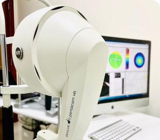
The Pentacam has become a tremendously valuable tool in assessing patients for laser vision correction. The Pentacam is a comprehensive eye scanner which provides data critical to the planning of the treatment.The Pentacam images the anterior segment of the eye by a rotating Scheimpflug camera measurement. This rotating process supplies pictures in three dimensions. The center of the cornea is measured very precisely because of this rotational imaging process. The measurement process lasts less than two seconds, and minute eye movements are captured and corrected simultaneously. By measuring 25,000 true elevation points, precise representation, repeatability and analysis are guaranteed.

Corneal Topography

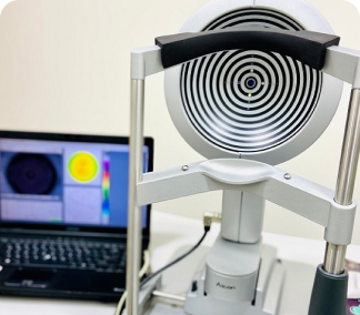
Corneal topography, also known as photokeratoscopy or videokeratography, is a non-invasive medical imaging technique for mapping the surface curvature of the cornea, the outer structure of the eye. Since the cornea is normally responsible for some 70% of the eye’s refractive power, its topography is of critical importance in determining the quality of vision and corneal Health

Optical Coherence Tomography (OCT)

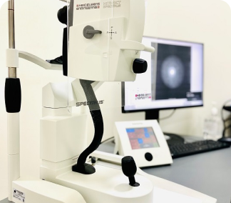
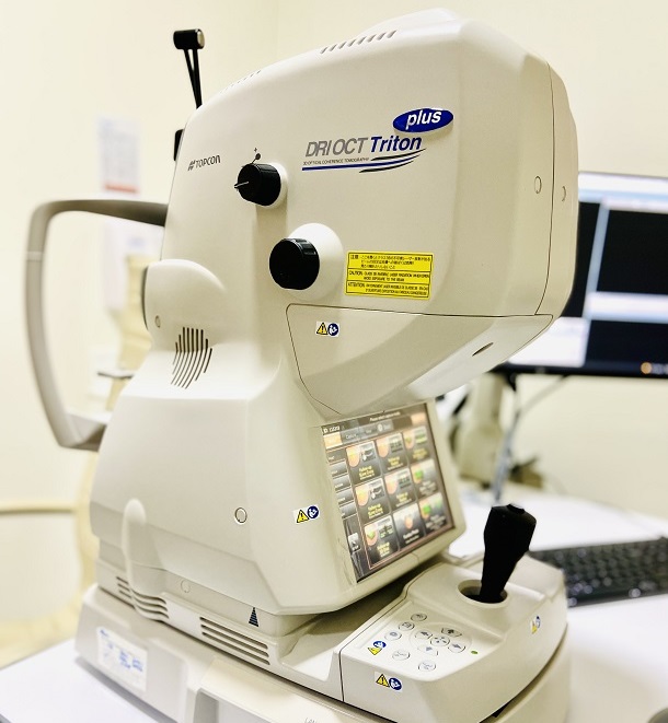
Optical coherence tomography (OCT) is a non-invasive imaging test. OCT uses light waves to take cross-section pictures of your retina.

With OCT, your ophthalmologist can see each of the retina’s distinctive layers. This allows your ophthalmologist to map and measure their thickness. These measurements help with diagnosis. They also provide treatment guidance for glaucoma and diseases of the retina. These retinal diseases include age-related macular degeneration (AMD) and diabetic eye disease.

Optical Coherence Tomography Angiography (OCTA)

Optical coherence tomography angiography (OCTA) is an imaging modality which can be applied in ophthalmology to provide detailed visualization of the perfusion of vascular networks in the eye. Compared to previous state of the art dye-based imaging, such as fluorescein angiography, OCTA is non-invasive, time-efficient, and it allows for the examination of retinal vasculature in 3D

Optical Coherence Tomography Angiography (OCTA)

Fluorescein Angiography

Fluorescein Angiography

(FA) is a diagnostic procedure that uses a special camera to record the blood flow in the RETINA – the light sensitive tissue at the back of the eye.

The test does not involve any direct contact with the eyes. Your eyes will be dilated before the procedure.

Fluorescein dye is injected into a vein in the arm/hand. As dye passes through the blood vessels of your eye, photographs are taken to record the blood flow in your retina. The photographs can reveal abnormal blood vessels or damage to the lining underneath the retina.

Fundus Photo

Color Fundus Retinal Photography uses a fundus camera to record color images of the condition of the interior surface of the eye, in order to document the presence of disorders and monitor their change over time.

A fundus camera or retinal camera is a specialized low power microscope with an attached camera designed to photograph the interior surface of the eye, including the retina, retinal vasculature, optic disc, macula, and posterior pole (i.e. the fundus).

The retina is imaged to document conditions such as diabetic retinopathy, age related macular degeneration, macular edema and retinal detachment.

Fundus Photo

Fundus photography is also used to help interpret fluorescein angiography as certain retinal landmarks visible in fundus photography are not visible on a fluorescein angiogram.

Your eyes will be dilated before the procedure. Widening (dilating) a patients pupil increases the angle of observation. This allows the technicians to image a much greater area and have a clearer view of the back of the eye.

Visual Field Test

Visual Field Test

A visual field test can determine if you have blind spots (called scotoma) in your vision and where they are. A scotoma’s size and shape can show how eye disease or a brain disorder is affecting your vision.

Visual field testing is an important part of regular eye care for people who are at risk for vision loss from disease and other problems.

People with the following conditions should be monitored regularly by their ophthalmologist, who will determine how often visual field testing is needed:

Glaucoma, Multiple sclerosis, Thyroid eye disease, Pituitary gland disorders, Central nervous system problems (such as a tumor that may be pressing on visual parts of the brain), and Stroke.

Long-term use of certain medications (such as Plaquenil, or hydroxychloroquine, which requires yearly visual field checkups)

People with diabetes and high blood pressure have a greater risk of developing blocked blood vessels in the optic nerve and retina. They may need visual field testing to monitor any effects of these conditions on their vision.
If your visual field is limited, your ability to drive may be in jeopardy. If you are concerned about vision loss or your ability to continue driving, talk with your ophthalmologist.

Pachymetry

Pachymetry is a simple, painless test to measure the thickness of your cornea — the clear window at the front of the eye. A probe called a pachymeter is gently placed on the front of the eye (the cornea) to measure its thickness. Pachymetry can help your diagnosis, because corneal thickness has the potential to influence eye pressure readings. With this measurement, your doctor can better understand your IOP reading and develop a treatment plan that is right for you. The procedure takes only about a minute to measure both eyes.

Pachymetry

A-Scan & B-Scan Tests

A-Scan & B-Scan Tests

What is an A-Scan?

An A-scan is short for amplitude scan. It is a type of ultrasound biometry that is provides data on the eye’s length. Determining eye length is crucial regarding many common sight disorders, including near-sightedness and far-sightedness. However, for these common conditions, traditional clinician exams are sufficient and far less invasive.

The A-scan provides data on the length of the eye, which is a major determinant in common sight disorders. The most common use of the A-scan is to determine eye length for calculation of intraocular lens power.

What is a B-Scan?

Similar to the A-scan, a B-scan (or brightness scan) is used in ophthalmology to produce a two-dimensional cross section of the eye and its orbit. It is most commonly used when a basic clinical examination isn’t revealing enough information.

Related Doctors

Patient Satisfaction Is Very Important To Us