


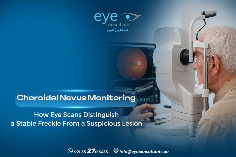
Choroidal nevus monitoring helps doctors follow a pigmented spot inside the eye and determine whether it remains stable over time. Most choroidal nevi are benign, but some may develop features that require closer evaluation. Baseline imaging and regular specialist follow-up provide a clear reference for detecting meaningful changes.
✆ WhatsappWhat Is a choroidal nevus monitoring and How Is It Usually Discovered?
Choroidal nevus monitoring begins with identifying the lesion and documenting its appearance. A choroidal nevus is usually a benign pigmented lesion in the choroid, often discovered during a routine dilated eye examination.
The ophthalmologist evaluates:
- Lesion size and thickness.
- Location within the eye.
- Pigmentation and appearance.
- Its relationship with the retina and optic nerve.
- Any associated retinal or subretinal changes.
An eye freckle monitoring plan may then be recommended according to the lesion’s characteristics.
Why Do Most Lesions Need a Baseline Record?
Most nevi remain stable, but changes in size or certain clinical features may increase concern. Choroidal nevus monitoring creates a baseline that can be compared with future examinations.
Depending on the case, documentation may include:
- Retinal photography.
- OCT imaging.
- Fundus autofluorescence.
- Ultrasound examination.
Using retinal photography follow-up can help doctors compare the lesion’s appearance over time, while other imaging methods provide additional structural information.
Which Scans Are Used to Monitor a Choroidal Nevus?
Different imaging techniques provide different types of information. Choroidal nevus monitoring may therefore involve more than one imaging method.
Choroidal nevus OCT can show the retinal layers and identify associated subretinal fluid. Fundus autofluorescence can help detect abnormal pigmentation, while ocular ultrasound for nevus can assess lesion thickness and internal characteristics.
The choice of imaging depends on the lesion and the findings during the eye examination.
Which Features Can Make a Nevus More Suspicious?
Choroidal nevus monitoring is particularly important when a lesion has features associated with a higher risk of growth or malignant transformation.
The ophthalmologist may pay closer attention to findings such as:
- Greater lesion thickness.
- Subretinal fluid.
- Orange pigment.
- Documented growth.
- Visual symptoms associated with the lesion.
- Other suspicious imaging characteristics.
These findings can help doctors assess nevus vs melanoma eye concerns and determine whether closer surveillance or specialist ocular-oncology assessment is appropriate.
What Should Patients Watch for Between Appointments?
Choroidal nevus monitoring should be combined with attention to new visual symptoms. A change in vision does not necessarily mean that a nevus has progressed, but it should not be ignored.
Seek ophthalmic assessment if you notice:
- New flashes or floaters.
- Unexplained blurred vision.
- New distortion in central vision.
- A noticeable change in visual quality.
The appropriate follow-up interval depends on the lesion’s appearance, imaging findings, and individual risk factors. (Watch this video)
Why Choose Eye Consultants Center?
Accurate retinal assessment depends on appropriate imaging and experienced interpretation. Eye Consultants Center has more than 14 years of ophthalmology services in Dubai, with doctors bringing over 30 years of clinical experience across complex retinal and vitreous conditions. The center provides advanced retinal evaluation supported by imaging such as fundus photography, OCT, and OCT-A.
The team includes Dr. Nihal Kenawy, Consultant Ophthalmologist in Medical Retina and Ocular Oncology, with over 20 years of experience and expertise in multimodal ocular-oncology imaging, including OCT, OCTA, FFA, and ICG.
If you have been diagnosed with a choroidal nevus, specialist follow-up can help establish an appropriate monitoring plan based on your individual findings. (Watch also)
Book your appointment at Eye Consultants Dubai for a comprehensive retinal and ocular-oncology assessment.
Contact us:
or visit Al Razi Building 64, Block C, 1st Floor, Unit 1017 – Dubai Healthcare City – Dubai – United Arab Emirates.
Working Hours
Monday to Thursday and Saturday: 8 AM to 8 PM
Friday: Closed
Sunday: 9 AM to 8 PM
Frequently Asked Questions
Can a choroidal nevus become melanoma?
A small proportion of choroidal nevi may develop into melanoma. This is why choroidal nevus monitoring and appropriate imaging are important for detecting concerning changes.
Does every choroidal nevus need treatment?
No. Most choroidal nevi are monitored rather than treated. The decision depends on the lesion’s appearance, size, imaging findings, and whether it changes over time.
How often should a choroidal nevus be checked?
There is no single schedule for every patient. The ophthalmologist determines follow-up according to the lesion’s characteristics and risk profile.
Can OCT detect changes in a choroidal nevus?
OCT can provide detailed information about the retina and identify associated changes such as subretinal fluid. It is usually interpreted alongside other examination and imaging findings.
Conclusion
Choroidal nevus monitoring provides a structured way to compare a lesion over time instead of relying on a single examination. Specialist assessment and appropriate imaging can help determine whether the lesion remains stable or requires closer evaluation.
✆ Whatsapp