


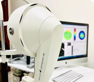
The Pentacam has become a tremendously valuable tool in assessing patients for laser vision correction. The Pentacam is a comprehensive eye scanner which provides data critical to the planning of the treatment..
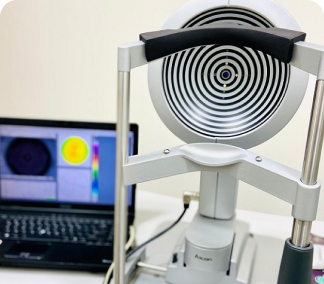
Corneal topography, also known as photokeratoscopy or videokeratography, is a non-invasive medical imaging technique for mapping the surface curvature of the cornea, the outer structure of the eye.
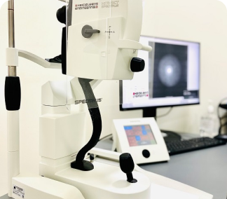
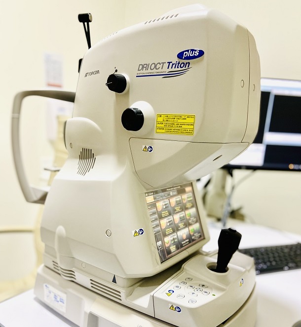
Optical coherence tomography (OCT) is a non-invasive imaging test. OCT uses light waves to take cross-section pictures of your retina.
Consultant Ophthalmologist
Vitreo Retinal & Anterior Segment Surgeon
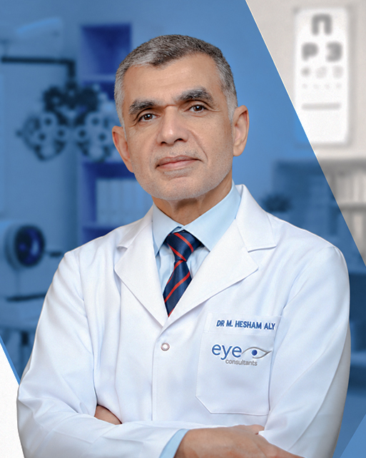
Consultant Ophthalmologist / Glaucoma & Cataract Surgeon
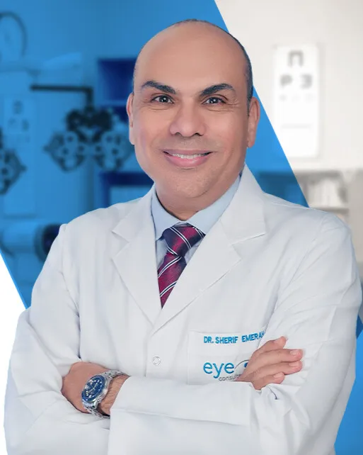
Consultant Ophthalmologist
Cornea And Refractive Surgeon
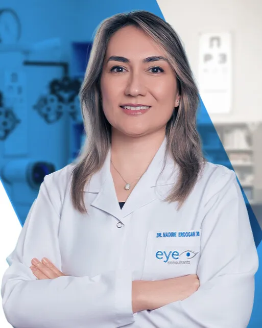
Consultant Ophthalmologist
Anterior Segment, Oculoplastic Surgeon & Pediatric Ophthalmology

Consultant Ophthalmologist
Cornea, Refractive And Anterior Segment Surgeon
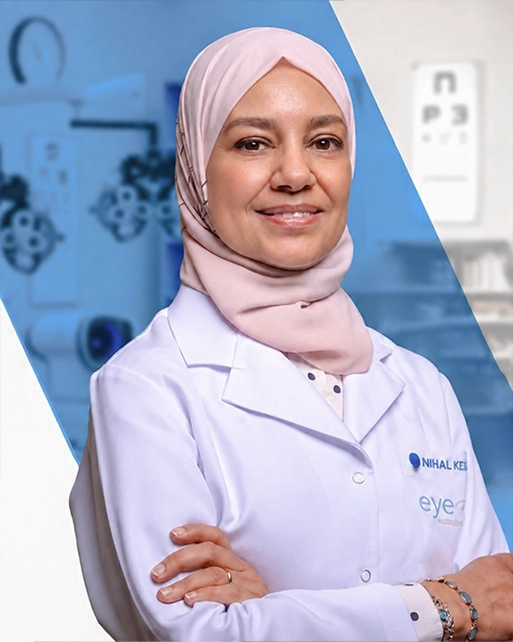
Consultant Ophthalmologist / Medical Retina, Ocular Oncology & Retinopathy of Prematurit
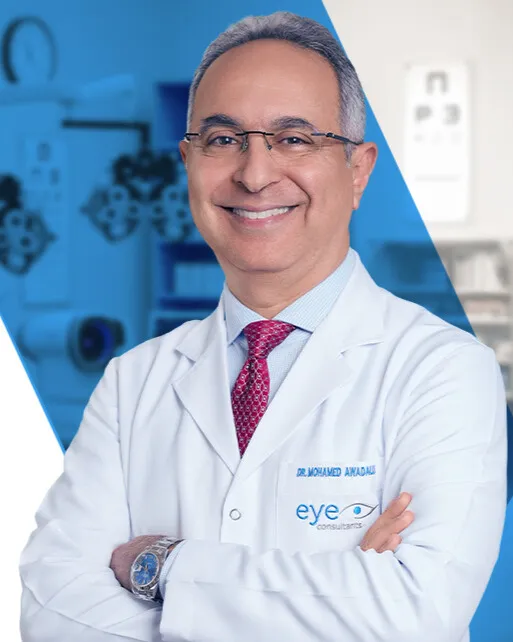
Consultant Ophthalmologist
Vitreo Retinal Surgeon (Visiting Doctor)
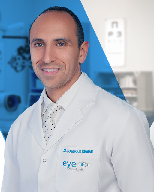
Specialist Ophthalmologist
Head Of Pediatric Ophthalmology & Strabismus Unit
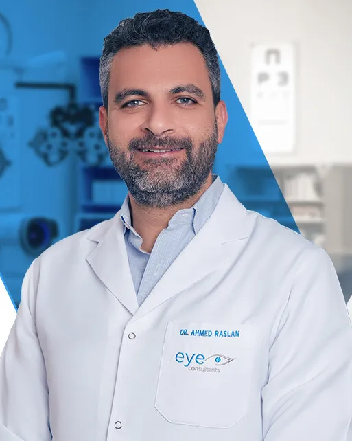
Specialist Ophthalmologist
Oculoplasty And Orbit Surgeon
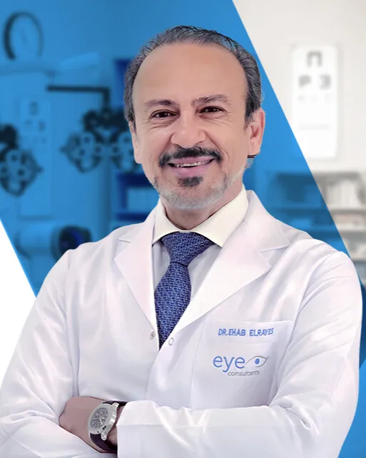
Consultant Ophthalmologist / Vitreo Retinal Surgeon