


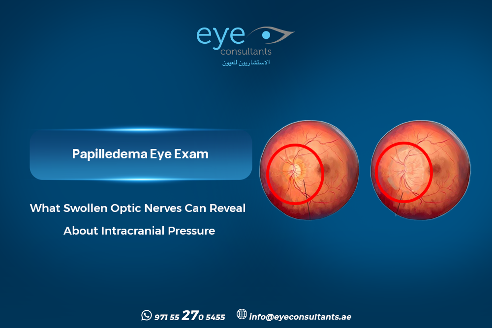
Papilledema eye exam can identify optic disc swelling that may be associated with increased pressure inside the skull, sometimes before major visual symptoms develop. Because an elevated optic disc can have several causes, careful assessment is important to determine whether true papilledema is present and whether further neurological evaluation may be needed.
What Does a Papilledema Eye Exam Reveal, and How Is It Different From Ordinary Optic-Nerve Swelling?
A papilledema eye exam begins with a detailed examination of the optic nerve and the patient’s visual function. Papilledema specifically refers to optic disc swelling caused by increased intracranial pressure, while other conditions can make the optic nerve appear elevated without true papilledema.
The examination may assess:
- Optic disc appearance and margins.
- Visual acuity and pupil responses.
- Retinal and optic-nerve changes.
- Visual-field function.
- Associated neurological symptoms.
A swollen optic nerve exam therefore focuses not only on identifying disc elevation, but also on understanding its possible cause.
Which Symptoms Can Make Optic Disc Swelling More Concerning?
Some patients with papilledema may initially have few or no visual symptoms. When symptoms occur, they can include headache, temporary visual dimming, nausea, vomiting, pulsatile tinnitus, or double vision.
The combination of headache and optic disc swelling may require particular attention, especially when symptoms are new, persistent, or worsening.
Other symptoms may include:
- Brief episodes of blurred or dim vision.
- New or persistent headaches.
- Double vision.
- Nausea or vomiting.
- Pulsing or whooshing sounds in the ears.
- Progressive visual changes.
These symptoms do not confirm papilledema by themselves, but they can help determine the urgency of further assessment.
Which Eye Tests Can Help Detect Papilledema?
Papilledema eye exam may involve several complementary investigations. Eye Consultants Center provides advanced ophthalmic investigations, including OCT and visual-field testing.
Depending on the clinical findings, assessment may include:
- Fundus examination and optic-nerve photography.
- OCT of the optic nerve and retinal nerve-fiber layer.
- Papilledema OCT to document structural changes.
- Automated perimetry, including a visual field for papilledema when clinically appropriate.
- Additional imaging when indicated.
The results should be interpreted together rather than relying on one test alone. Imaging, visual function, symptoms, and medical history all contribute to the assessment.
How Can Doctors Distinguish Papilledema From Pseudopod pill edema?
An elevated optic disc does not always indicate increased pressure inside the skull. Optic disc drusen, for example, can create pseudopod pill edema and may resemble true disc swelling.
Certain raised intracranial pressure eye signs can help raise clinical concern, but additional testing may be required when the appearance is uncertain.
The ophthalmologist may use:
- Enhanced-depth OCT.
- Fundus autofluorescence.
- B-scan ultrasonography.
- Optic-nerve photography.
- Comparison with previous examinations.
These tests provide complementary information and can help distinguish true optic disc edema from other causes of an elevated-looking nerve.
When Is Urgent Neurological Evaluation Required?
Papilledema eye exam findings may sometimes indicate increased intracranial pressure that requires prompt medical investigation. Potential causes include idiopathic intracranial hypertension, intracranial masses, and cerebral venous disorders.
Depending on the findings, further evaluation may involve brain imaging and neurological assessment.
Prompt medical evaluation is particularly important when there is:
- Rapidly worsening vision.
- Severe or persistent headache.
- New double vision.
- New neurological symptoms.
- Significant visual-field changes.
The appropriate next step depends on the examination and the suspected underlying cause. (Watch also)
Why Choose Eye Consultants Center?
Accurate optic-nerve assessment requires appropriate diagnostic testing and experienced interpretation. Eye Consultants Center provides advanced ophthalmic investigations in Dubai, including OCT, OCT-A, fundus imaging, and visual-field testing. The center has more than 14 years of experience, while its doctors bring over 30 years of clinical expertise across ophthalmic specialties. (Watch this video)
The medical team includes experienced ophthalmologists and vitreoretinal specialists involved in the assessment of complex retinal and optic-nerve conditions.
If an abnormal optic disc has been identified, specialist assessment can help determine whether further ophthalmic or neurological evaluation is appropriate.
Book your appointment at Eye Consultants Dubai for a comprehensive optic-nerve and retinal assessment.
Contact us:
or visit Al Razi Building 64, Block C, 1st Floor, Unit 1017 – Dubai Healthcare City – Dubai – United Arab Emirates.
Working Hours:
Monday to Thursday and Saturday: 8 AM to 8 PM
Friday: Closed
Sunday: 9 AM to 8 PM
FAQ’S About Papilledema Eye Exam
Can papilledema occur without obvious symptoms?
Yes. Early papilledema may cause few noticeable symptoms. This is why an abnormal optic-disc appearance should be assessed even when vision seems relatively normal.
Does optic disc swelling always mean increased intracranial pressure?
No. Conditions such as optic disc drusen can create Pseudopod pill edema without increased intracranial pressure. Clinical examination and appropriate imaging help distinguish these conditions.
Can OCT confirm papilledema by itself?
No. OCT provides valuable structural information, but it should be interpreted alongside the clinical examination, symptoms, visual fields, and other investigations when appropriate.
What should I do if swollen optic nerves are found?
A papilledema eye exam can help clarify the cause of optic-disc elevation. Depending on the findings and symptoms, the ophthalmologist may recommend additional eye testing or neurological evaluation.
Conclusion
Papilledema eye exam is more than a routine vision check. Careful optic-nerve examination and appropriate investigations can help identify concerning findings and determine whether further evaluation is required.