


Gonioscopy for glaucoma diagnosis can reveal an important part of glaucoma risk that an eye pressure reading cannot show: the anatomy of the eye’s drainage angle. A normal pressure measurement does not automatically mean the angle is open or that glaucoma has been ruled out. Understanding the angle helps distinguish different glaucoma patterns and guides the next stage of assessment.
Gonioscopy for Glaucoma Diagnosis What Is the Drainage Angle and Why Does It Matter?
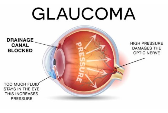
Gonioscopy for glaucoma diagnosis examines the anterior chamber angle, where fluid drains from the eye. If the angle is narrow or closed, fluid outflow can become restricted, changing the risk of angle closure. Gonioscopy is the standard clinical method for directly assessing this area.
- Shows whether the drainage angle is open, narrow, or closed.
- Helps identify abnormal structures or adhesions.
- Can help distinguish different types of glaucoma.
- Provides information that routine tonometry cannot provide.
This makes a Glaucoma Evaluation useful when a more complete assessment is needed.
Open Angle vs Narrow Angle: What Is the Difference?
Glaucoma does not follow one single pattern. Open angle disease can damage the optic nerve while the drainage angle remains open, while angle closure involves narrowing or closure of this drainage pathway.
- Open angle glaucoma often develops gradually and may have few early symptoms.
- Narrow or occludable angles may carry a risk of angle closure.
- Gonioscopy can reveal the anatomy behind an unexpected pressure reading.
- It can also identify findings associated with secondary glaucoma.
This distinction helps the ophthalmologist choose the appropriate glaucoma workup rather than relying on pressure alone.
What Happens During Gonioscopy?
During an eye angle exam, the doctor uses a special contact lens after applying topical anesthetic. The lens allows the drainage angle to be viewed through a slit lamp because the angle cannot normally be seen directly through the cornea.
- The patient looks in specific directions while the lens is gently positioned.
- The doctor examines different areas of the angle.
- Gentle indentation may be used when necessary.
- The examination can reveal narrowing, pigmentation, abnormal blood vessels, or adhesions.
The procedure is brief, does not involve surgery, and is generally well tolerated.
How Does Gonioscopy Complement OCT, Visual Fields, and Eye Pressure?
Gonioscopy for glaucoma diagnosis answers a structural question that other tests do not answer in the same way. Tonometry measures intraocular pressure, OCT evaluates the optic nerve and retinal nerve fiber layers, while visual field testing assesses functional vision.
- Tonometry: measures eye pressure.
- OCT: evaluates structural changes in the optic nerve and retinal layers.
- Visual field testing: identifies areas of reduced visual sensitivity.
- Gonioscopy: assesses the drainage angle and helps classify glaucoma.
Together, these findings create a more complete picture of the patient’s condition.
When Might a Narrow Angle Need Laser or Closer Monitoring?
Gonioscopy for glaucoma diagnosis can identify eyes with sufficiently narrow angles to require closer assessment. Management depends on the angle anatomy, symptoms, pressure, optic nerve status, and evidence of previous or ongoing angle closure.
- Some eyes may require closer monitoring.
- Laser peripheral iridotomy may be considered for selected angle closure mechanisms.
- Sudden severe eye pain, blurred vision, halos, headache, nausea, or vomiting requires urgent assessment.
A narrow angle does not automatically mean that glaucoma is present.
Why Choose Eye Consultants Center?
At Eye Consultants, glaucoma assessment is supported by a dedicated glaucoma service and a broader diagnostic pathway. The center provides tonometry, OCT, and visual field testing, while its medical team includes consultants with specific glaucoma and cataract expertise.(Watch this video)
- Comprehensive glaucoma assessment.
- Advanced diagnostic imaging.
- Specialized glaucoma expertise.
- Follow-up based on clinical findings and comparison over time.
Book your consultation at Eye Consultants in Dubai for a comprehensive glaucoma evaluation or a glaucoma second opinion.
Contact us
or visit Al Razi Building 64, Block C, 1st Floor, Unit 1017 – Dubai Healthcare City – Dubai – United Arab Emirates.
Working Hours:
Monday to Thursday and Saturday: 8 AM to 8 PM
Friday: Closed
Sunday: 9 AM to 8 PM
Frequently Asked Questions
Can normal eye pressure rule out glaucoma?
No. Glaucoma can occur even when pressure readings are within the statistically normal range, so other parts of the examination remain important.
Is gonioscopy painful?
Most patients experience little more than mild pressure. A topical anesthetic is applied before the lens is positioned.
Is gonioscopy only used for narrow angle glaucoma?
No. It is also important in assessing open angle glaucoma and identifying secondary causes affecting the drainage angle.
Can gonioscopy replace OCT or visual field testing?
No. Each test provides different information, and they are often interpreted together.
Conclusion
Gonioscopy for glaucoma diagnosis provides information that routine pressure measurement cannot provide. By examining the drainage angle and combining its findings with OCT, visual field testing, and pressure measurements, the ophthalmologist can build a more complete picture of glaucoma risk and determine the appropriate next step.